Spectacular Winning Photos Of The 2018 Nikon Small World Contest Reveal The Hidden Beauty Of A Microscopic World
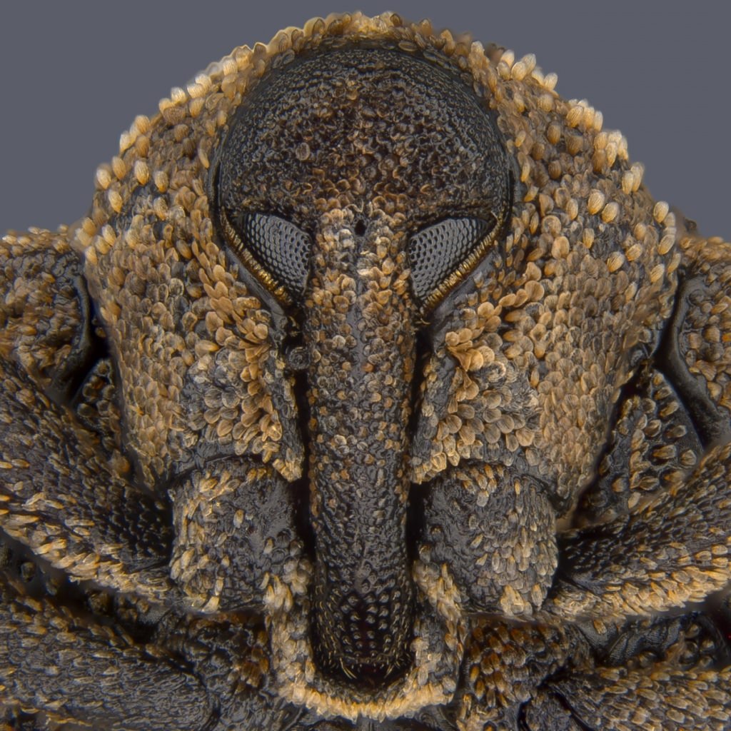
Eye of a “Metapocyrtus subquadrulifer” beetle, Yousef Al Habshi

For the 44th year, Nikon celebrates the invisible world by organizing Small World Photomicrography Competition. As imaging and microscope technologies evolve, scientists, professional photographers continue to push the boundaries of micrograph. This year over 2500 scientists and artist from 89 countries submitted their work.
After being evaluated on originality, informational content, technical proficiency, and visual impact, Yousef Al Habshi from Abu Dhabi was declared the winner. His incredible image of an Asian Red Palm weevil’s eye is a close up look at the insect’s striking anatomy.
More: Nikon Small World, Facebook, Instagram
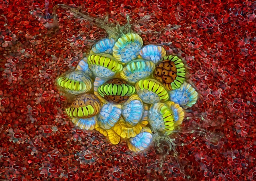
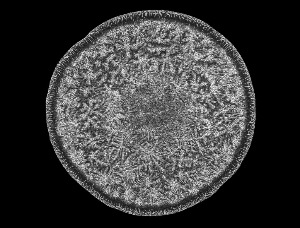
Fern sorus” (structures producing and containing spores), Rogelio Moreno
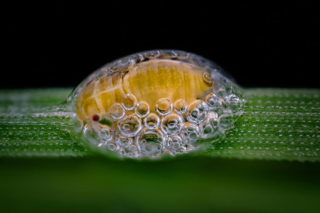
Spittlebug nymph in its bubble house, Saulius Gugis
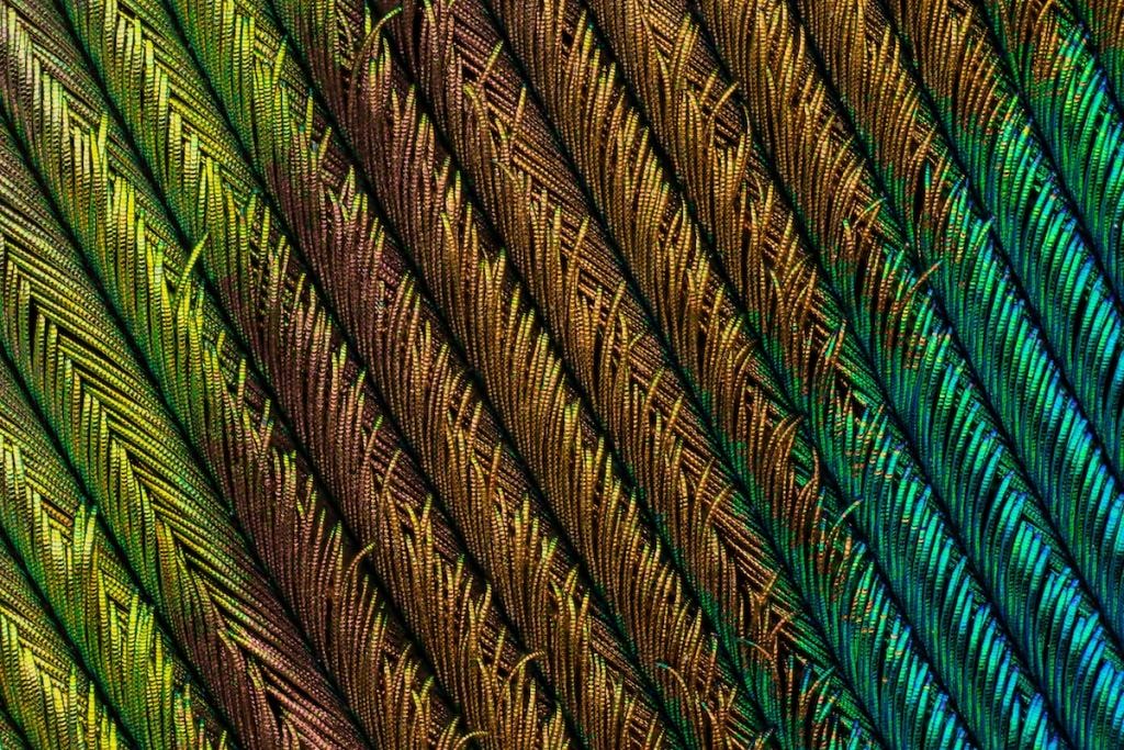
Peacock feather section, Can Tunçer
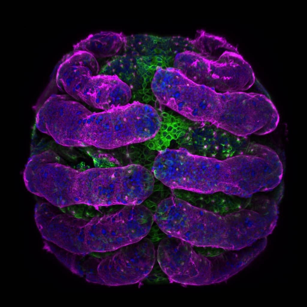
“Parasteatoda tepidariorum” (spider embryo) stained for embryo surface (pink), nuclei (blue) and microtubules (green), Dr. Tessa Montague
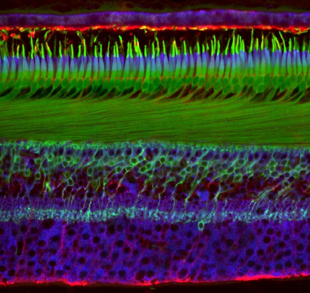
Primate foveola (central region of the retina), Hanen Khabou
Human tear drop, Norm Barker
Portrait of “Sternochetus mangiferae”, Pia Scanlon
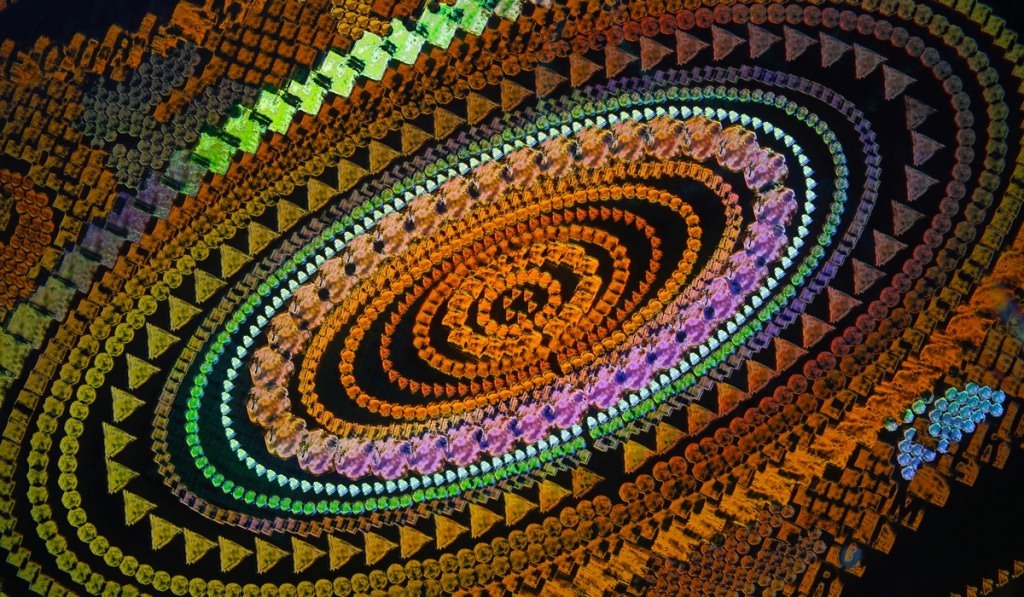
Security hologram, Dr. Haris Antonopoulos
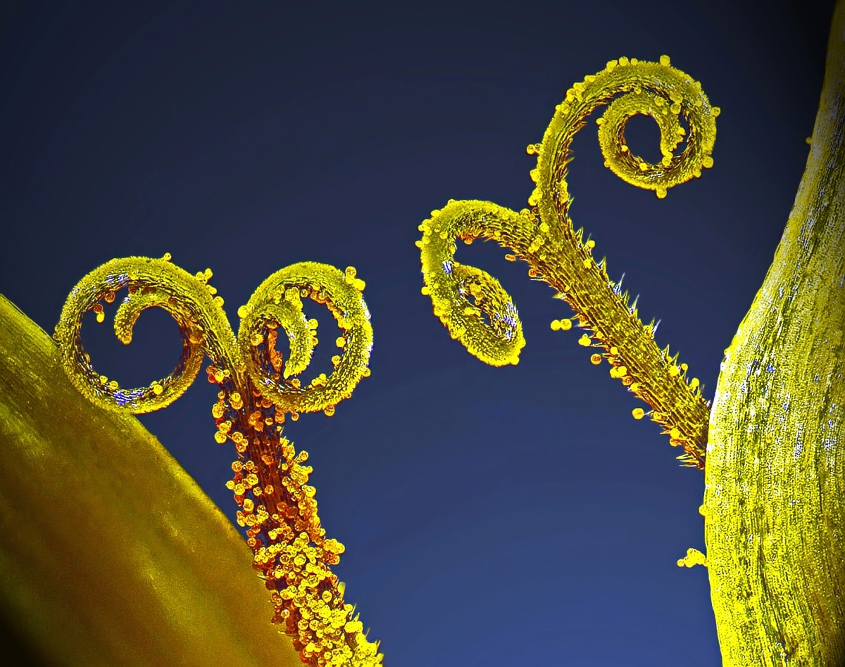
Stalks with pollen grains, Dr. Csaba Pintér
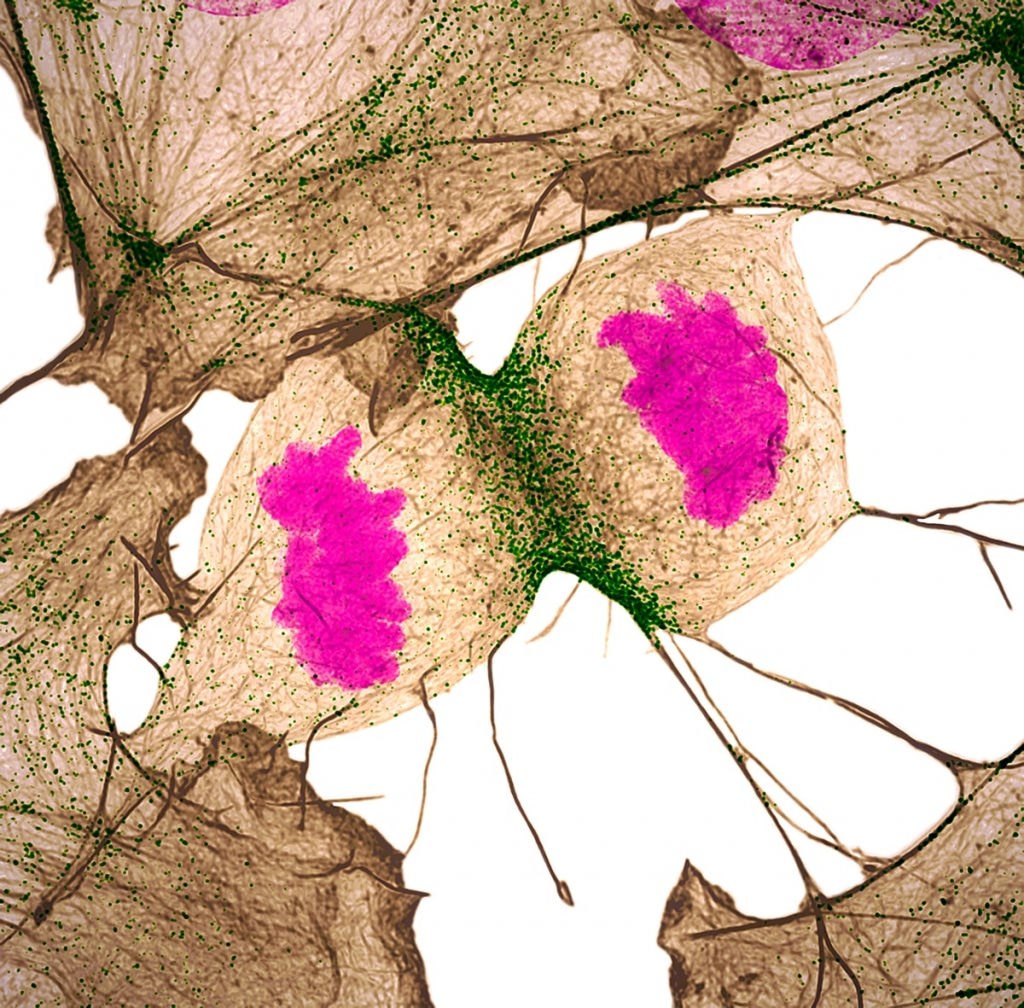
Human fibroblast undergoing cell division, showing actin (gray), myosin II (green) and DNA (magenta), Nilay Taneja & Dr. Dylan Burnette
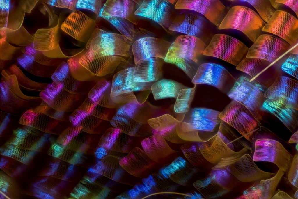
Urania ripheus” (butterfly) wing scales, Luciano Andres Richino
“Balanus glandula” (acorn barnacle), Charles Krebs
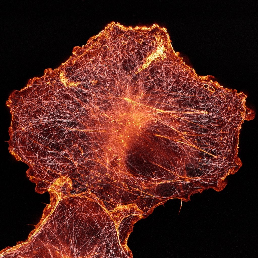
African green monkey cell (COS-7) stained for actin and microtubules, Andrew Moore & Dr. Erika Holzbaur
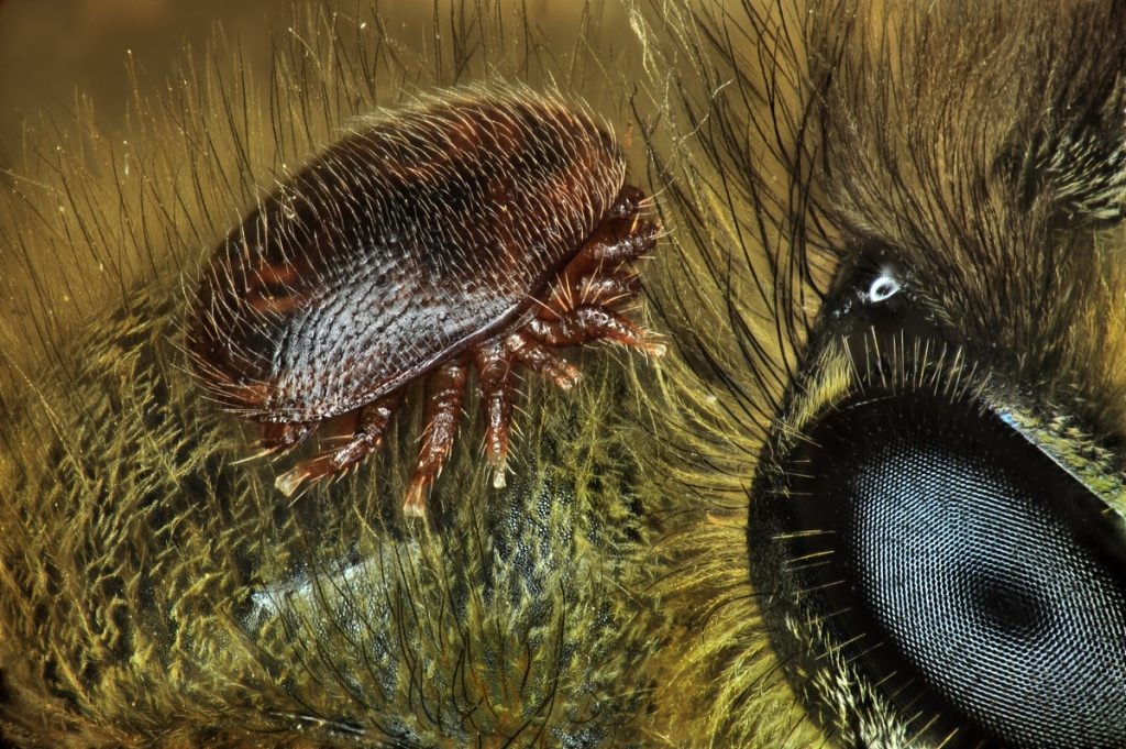
“Varroa destructor” (mite) on the back of “Apis mellifera” (honeybee), Antoine Franck
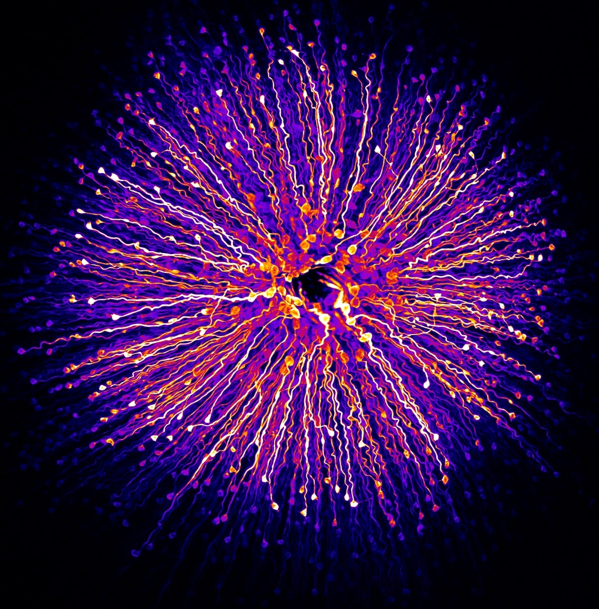
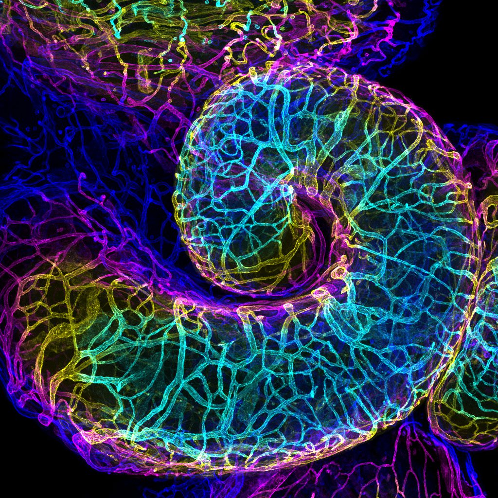
Mouse oviduct vasculature, Dr. Amanda D. Phillips Yzaguirre
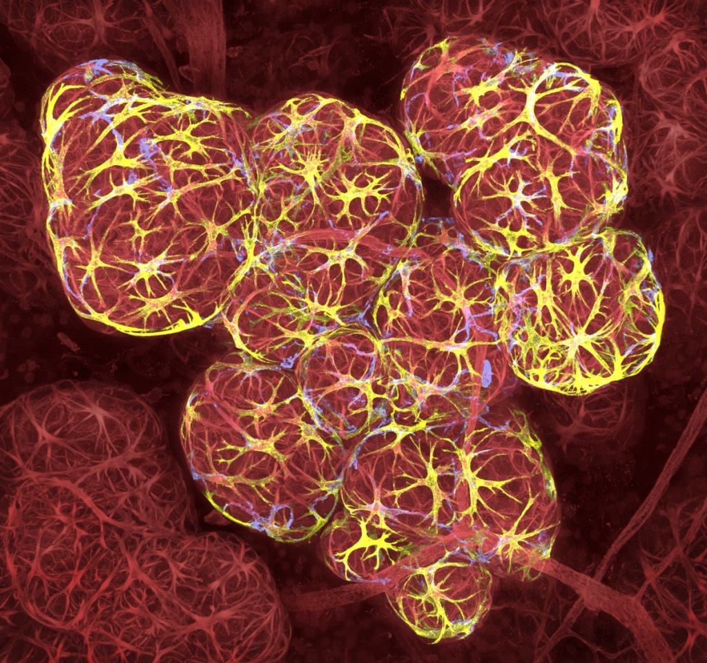
Breast tissue in lactation: Milk filled spheres (red) surrounded by tiny muscle cells that squeeze out milk (yellow) and immune cells that monitor for infection (blue), Caleb Dawson
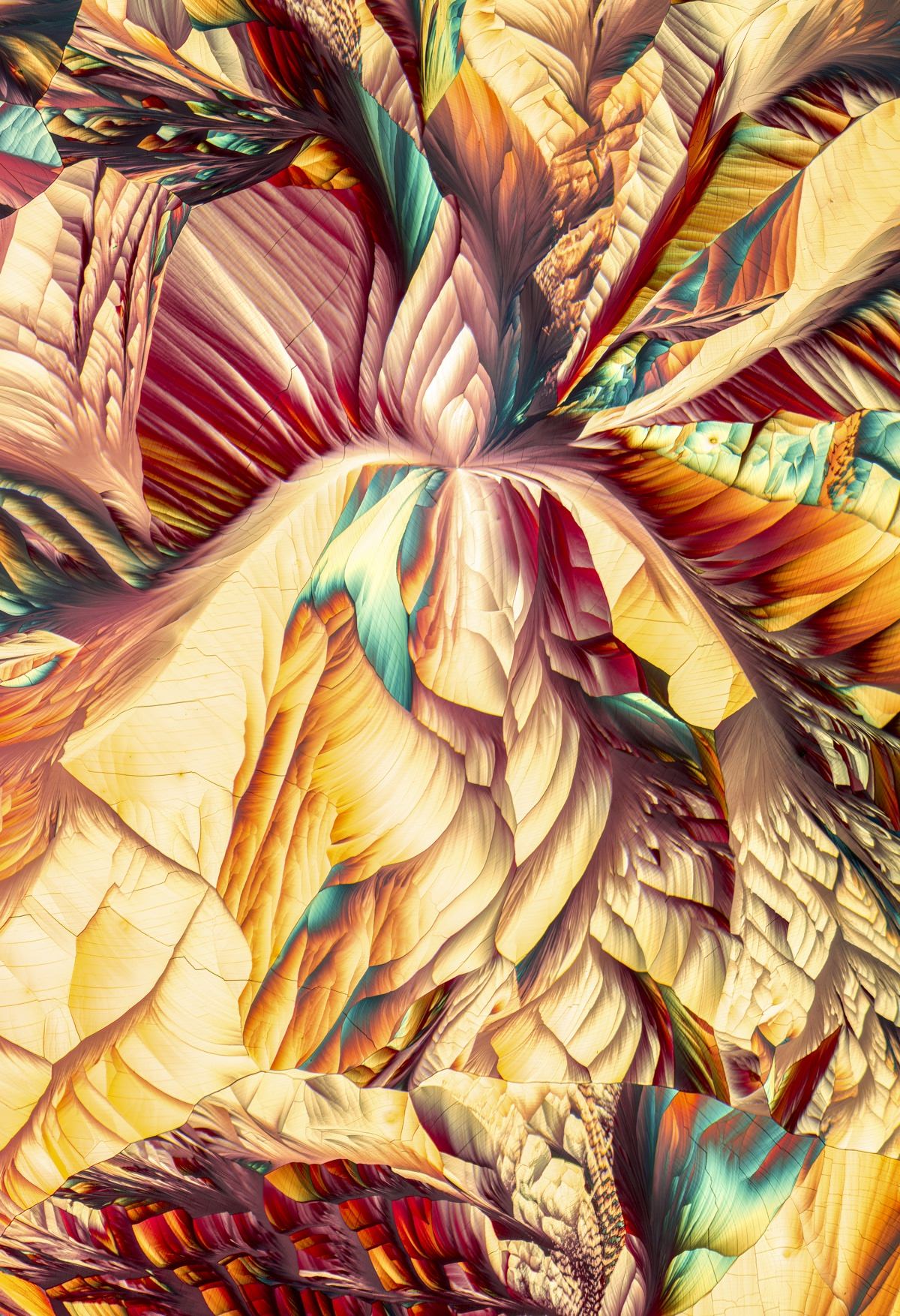
Amino acid crystals (L-glutamine and beta-alanine), Justin Zoll
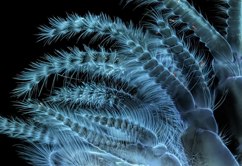
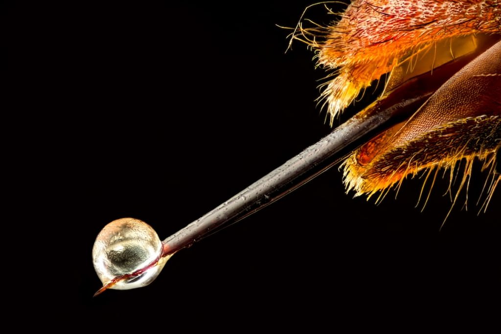
“Vespa velutina” (Asian hornet) with venom on its stinger, Pierre Anquet
Human retina, Dr. Nicolás Cuenca & Isabel