Magnificent Microcosms: Captivating Images of Imperiled Insects
The Rocky Mountain locust
Billions of Rocky Mountain Locusts, known as Melanoplus spretus, used to flock to the Great Plains in large numbers until the late 1800s. After this, the swarms stopped appearing and the locusts have not been seen since 1902.
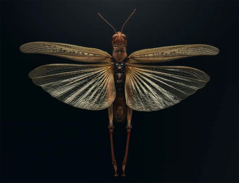
In an effort to raise awareness of the beauty of insects, British photographer Levon Biss has created an exhibition, Extinct & Endangered: Insects in Peril, showcasing stunning photographs taken with microscope lenses. His work is a reminder of how we often don’t appreciate the beauty of such creatures until it’s too late – a stark reality that’s seen in the fate of the once prolific Rocky Mountain Locust (Melanoplus spretus), last seen in 1902. Biss’ photographs, made from up to 10,000 individual images, captures the intricate details of his subject matter, drawing attention to the importance of preserving the planet’s biodiversity. Continue reading »
Polish Scientist’s Incredible Photographs of Microscopic Creatures
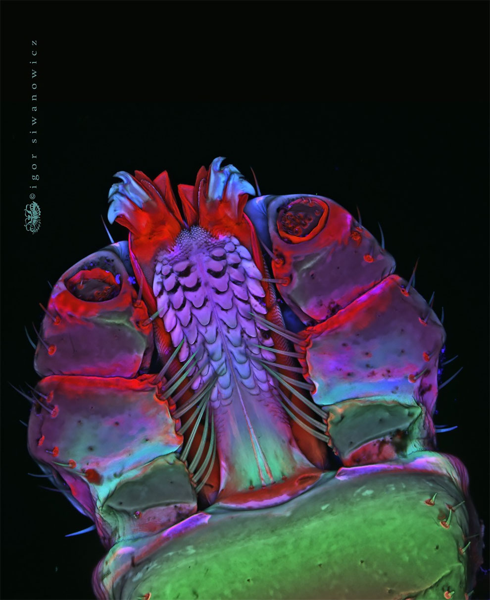
Photographer Igor Siwanowicz took these incredible photographs of tiny creatures. Dr Siwanowicz, a neurobiologist at the Howard Hughes Medical Institute’s Janelia Farm Research Campus, Virginia, shows us the wonders of life in kaleidoscopic color. Continue reading »
Art in A Petri Dish: The Agar Art Awards 2020
Scientists from around the world submitted art grown in petri dishes for the American Society of Microbiology’s annual contest, which has announced the winners. Restricted access to labs broadened the remit, with traditional art on the beauty of microbes accepted for the first time.
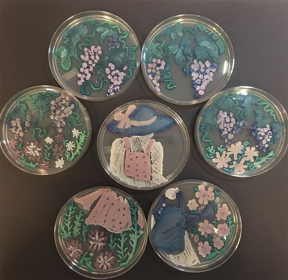
First place in the traditional general category was awarded to The Gardener by Joanne Dungo, from Northridge Hospital Medical Center in Northridge, California. Continue reading »
The Surprising Beauty Of Microscopic Insects, Isopods And Crustaceans Illuminated With Polarized Light
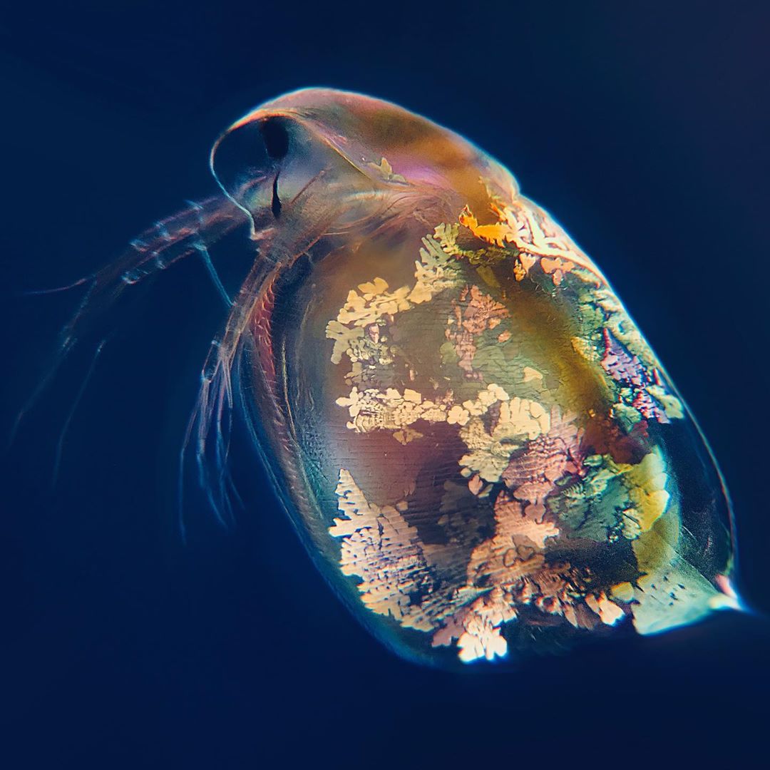
Biomedical scientist and microscopy enthusiast My Microscopic World created a really beautiful explanatory video that reveals the surprising beauty of various forms of tiny life as shown under a microscope that has been adapted with polarized life. These micro forms of life include insect larvae, isopods and crustaceans. Continue reading »
Nikon Small World 2019: The Best Microscope Photos Of The Year
Nikon has announced the winners of the 2019 Small World Photomicrography Competition, and has once more shared some of the winning and honored images with us. The contest invites photographers and scientists to submit images of all things visible under a microscope. This year, first place was awarded to Teresa Zgoda and Teresa Kugler for their painstakingly prepared photo of a turtle embryo, using fluorescence and stereo microscopy. More than 2,000 entries were received from 100 countries in 2019, the 45th year of the competition.
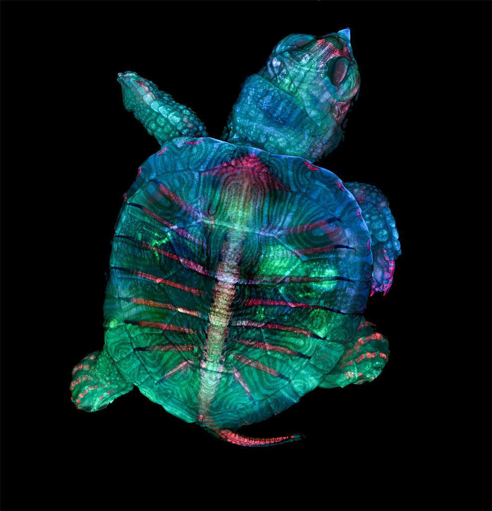
1st Place: Teresa Zgoda & Teresa Kugler, Campbell Hall, New York, USA. Fluorescent turtle embryo. Stereomicroscopy, Fluorescence, 5x (Objective Lens Magnification). (Photo by Teresa Zgoda/Nikon’s Small World 2019) Continue reading »
Here’s What Marijuana Looks Like Under The Microscope
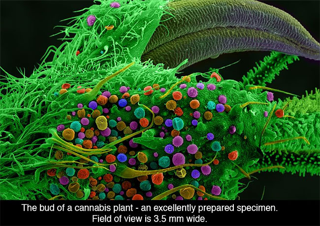
Now you can see how cannabis appears to the scientists who study it, thanks to a book called ‘Cannabis Under The Microscope: A Visual Exploration of Medicinal Sativa and C. Indica’ by Ford McCann. The book features over 170 images of cannabis in its full glory, taken with optical and scanning electron microscopes. Continue reading »
This 3D Printed Coffee Table Shows Art As Seen Under The Microscope
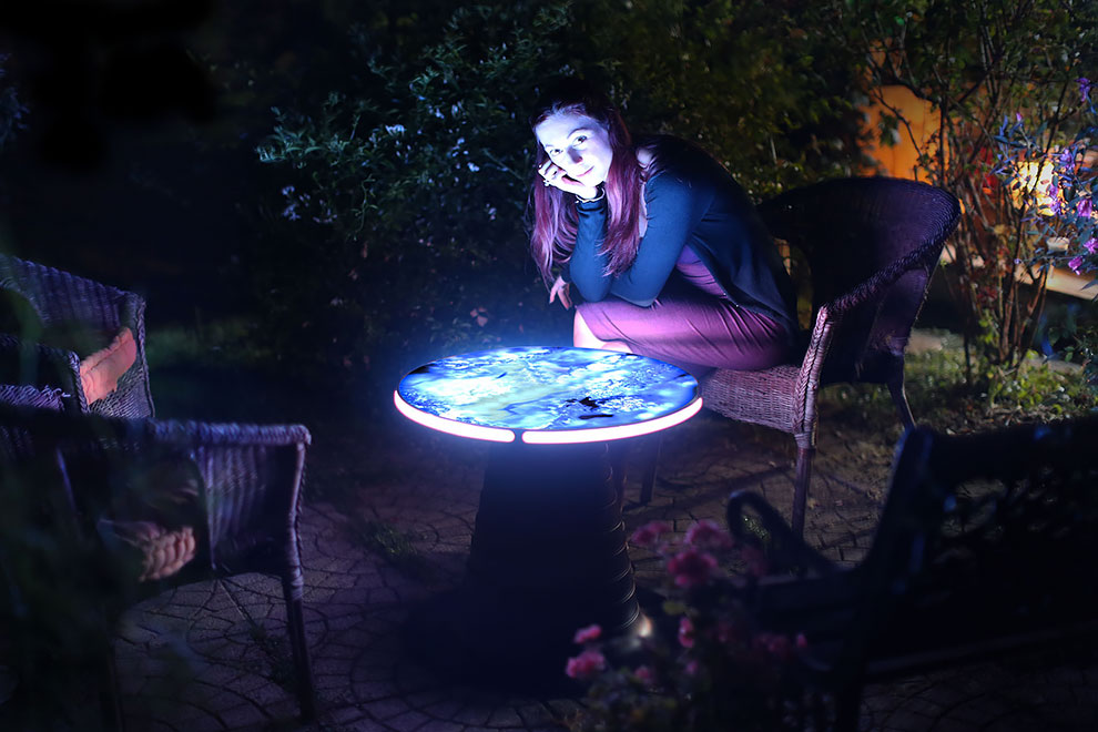
Scientists have long been fascinated with viewing objects under the microscope, and it’s easy to see why. You get a fresh perspective on everyday items that look completely different under the lens. With pretty patterns and vivid colors, there’s a sense of surprise when you get a closer look at images that can’t be seen with the naked eye. Continue reading »
Microsculpture: Insect Portraits Under The Microscope By Levon Biss
Jewel Longhorned Beetle
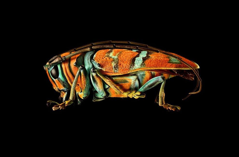
Levon Biss is a talented British photographer and filmmaker, who’s typically shoots portraits of world-class athletes. His passion for nature led him to create the striking “Microsculpture” project, a unique photographic study of insects in mind-blowing magnification. His talent for capturing insects started as a side-project in his home, and featured bugs caught by his son, and now Levon embraced the world of macrophotography and has taken the genre to a new level. Continue reading »
Everyday Things That Look Totally Different Under A Microscope
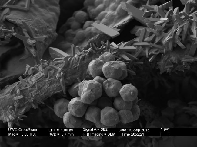
All the common objects are kinda boring when you look at them, but the situation changes when an awesome Electron Microscope comes on the scene. A scanning electron microscope (SEM) is a type of electron micrograph that makes images of a sample by scanning it with a focused beam of electrons. These are the most amazing images of what is too small to see with the naked eye. Here: Ground black pepper Continue reading »
This Is What Peacock Feathers Look Like Under A Microscope
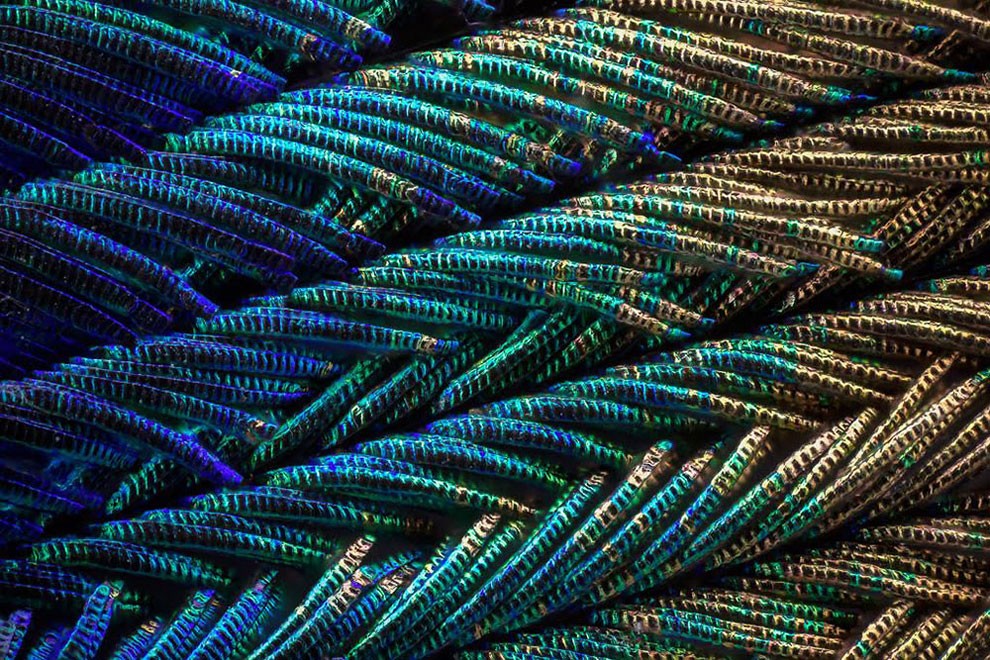
Canadian software engineer and photographer Waldo Nell shows us just how fabulous peacock feathers are – even at a microscopic level. Using an Olympus BX 53 microscope, Nell employed a technique called ‘photo stacking’. It involves combining hundreds of pictures taken at different focal points to get an image with a greater depth of field. Continue reading »
Alien-like Flowers Seen Under the Microscope
These images have been created using a colour scanning electron microscope (SEM) by the award-winning Eye of Science, comprised of snapper Oliver Meckes and biologist Nicole Ottawa. For a decade the pair, based in Reutlingen in the south of Germany, worked with an old SEM they saved from the scrapheap, but for the last five years they have used a £250,000 FEI Quanta Series Field Emission SEM. Oliver said: “Flowers are beautiful in ‘normal’ view, but when you look closer, some parts get very bizarre and unexpected structures appear – flowers within flowers, worlds within worlds”.
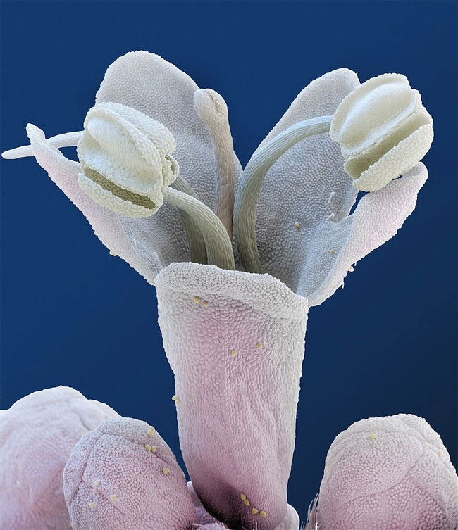
A Valerian flower as viewed under a coloured scanning electron microscope. (Photo by Oliver Meckes/Barcroft Media)
Continue reading »