The Surprising Beauty Of Microscopic Insects, Isopods And Crustaceans Illuminated With Polarized Light – Design You Trust

The Surprising Beauty Of Microscopic Insects, Isopods And Crustaceans Illuminated With Polarized Light

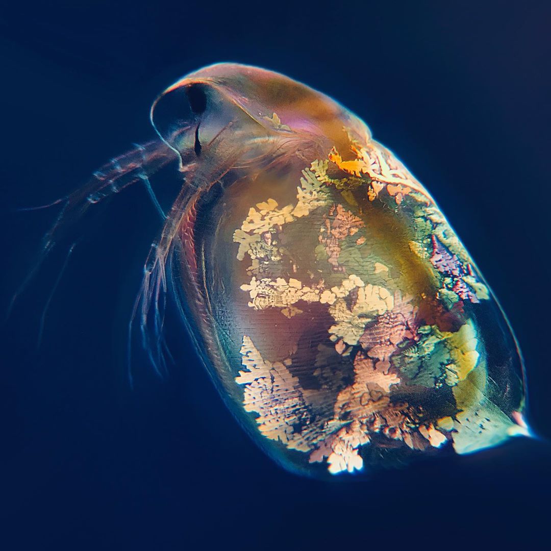
Biomedical scientist and microscopy enthusiast My Microscopic World created a really beautiful explanatory video that reveals the surprising beauty of various forms of tiny life as shown under a microscope that has been adapted with polarized life. These micro forms of life include insect larvae, isopods and crustaceans.

More: Instagram h/t: laughingsquid

“A few weeks ago, I modified my microscope to be able to use polarized light to illuminate my samples. This gives some incredibly beautiful and alien-like footage, so I of course made a video with this technique.”

View this post on Instagram

This is a crane fly, or a tipula if you are fancy. These are found all over the world and look a bit like a huge mosquito. But in contrast to the mosquito, crane flies can’t bite or sting. Some people refer to these insects as “mosquito hawks”, which is a bit misleading as the crane fly is incapable of eating other insects. If the adult crane fly feeds at all during its life, if feeds on nectar. . These images are as always taken with my iPhone mounted on my microscope with a @ilabcam . The colorful background is not photoshop, it is made optically with polarized light! . #science #biology #microscopy #entomology #labcam #shotoniphone #insect #cranefly #tipula #ecology #bug #macrophotography #macro #extrememacro #microscope #closeup #magnification #facts #nature #animal

A post shared by My Microscopic World (@my.microscopic.world) on

View this post on Instagram

This is not an alien world filled with giants, but a tiny green spec of plant material. How many tardigrades can you spot? . NARRATION: “This tiny piece of plant material is about the same size as a grain of sand and as you can see, this microworld has a few inhabitants. The two big ones are mites, and if you look closely, you are able to see several tardigrades waddling around. Luckily for the tardigrades, these mites are herbivores and won’t harm them. Mites are very common in nature, both on land and in water, and despite what you might think, most species are harmless to humans and only a few are able to cause disease. Most mites are under a millimeter in size and was first studied in detail under a microscope in 1665 by Robert Hooke who described them as “very prettily shaped insects”. But mites are actually not insects, but arachnids, which means that they are closely related to spiders and scorpions!” . If you enjoyed the music in this video and want to hear more, I highly recommend that you go check out the talented artist @animamusic1 as well as his SoundCloud (link in his bio). . This was as always filmed with my iPhone mounted on my microscope with a @ilabcam . . #science #biology #microscopy #labcam #shotoniphone #underthemicroscope #tardigrade #nature #life #microworld #microcosmos #ecology #waterbear #microscope #closeup #magnification #scienceandtechnology #invertebrate #narratedpost #mite #microscopic #animal #mosspiglet #bug #biologia #arachnid #arthropod #facts

A post shared by My Microscopic World (@my.microscopic.world) on

View this post on Instagram

Let’s try something new. As you can hear I have narrated this post and if you would like to see more of these or think that I should just shut up and do as I have always done, let me know in the comments. . NARRATION: “This is a bunch of tardigrades, and I believe these are from the genus milnesium. They are predatory tardigrades feeding on single celled organisms, microscopic animals or even on other smaller tardigrades. These ones are very big and fast compared to other species of tardigrades, in fact they were so big that I could see them waddling around on the slide before I put it under the microscope. I isolated the tardigrades from a small piece of moss I found on a bench, so the next time you sit down outside you might have quite a few of them crawling around on your bud looking for a meal.” . If you enjoyed the music in this video and want to hear more, I highly recommend that you go check out the talented artist @animamusic1 as well as his SoundCloud (link in his bio). . This was as always filmed with my iPhone mounted on my microscope with a @ilabcam . . #science #biology #microscopy #labcam #shotoniphone #underthemicroscope #tardigrade #nature #life #microworld #microcosmos #ecology #waterbear #microscope #closeup #magnification #scienceandtechnology #invertebrate #narratedpost #milnesium #microscopic #animal #mosspiglet #bug #biologia #predator

A post shared by My Microscopic World (@my.microscopic.world) on

View this post on Instagram

This is the larva of a hoverfly, or syrphidae if you are fancy. The larva has been magnified over 100 times and inside of the animal you are able to see a bunch of white tubes. This network is actually the larva’s respiratory system which it, unsurprisingly, uses to breath. And just like your windpipe, these tubes are called trachea (plural tracheae). The biggest parts of the tubes are connected to pores on the outside of the body called spiracles. As the tubes moves towards the center of the animal, they branch of into smaller and smaller tubes which finally reach a tissue/cells that needs oxygen. . If you “enjoyed” the scary music in this video and want to hear more, I highly recommend that you go check out the talented artist @animamusic1 as well as his SoundCloud (link in his bio). . This was as always filmed with my iPhone mounted on my microscope with a @ilabcam . . #science #biology #microscopy #labcam #shotoniphone #underthemicroscope #nature #life #microworld #microcosmos #ecology #entomology #microscope #closeup #magnification #scienceandtechnology #insects #hoverfly #trachea #breathing #larvae #animal #larva #respiratorysystem #bug #biologia

A post shared by My Microscopic World (@my.microscopic.world) on

View this post on Instagram

Today I took a drop of my sample water, added it to a slide and put a coverslip on top like I usually do. But then I noticed something I hadn’t paid attention to before; how beautiful bubbles look under the microscope! So, I decided to take an artistic break and take a few nice photos of some bubbles! . All the images are magnified over 150 times and are as always taken with my iPhone mounted on my microscope with a @ilabcam adapter. . #science #biology #microscopy #bubbles #labcam #shotoniphone #underthemicroscope #art #artistic #microworld #microcosmos #phisics #microscope #closeup #magnification #scienceandtechnology #beautiful #polarizedlight #photography #trippy #soap #soapbubbles

A post shared by My Microscopic World (@my.microscopic.world) on

View this post on Instagram

This is a larva of a marine snail, or a veliger of a gastropod if you are fancy. The rapid beating hair-like projections are called cilia and the animal uses them to swim through the water and to feed. The cilia create a current of water and food particles towards its mouth to be ingested. If you look closely in one of the last shots, you are able to see food particles being swirled around inside the stomach which aids in digestion. You are also able to see a beating heart inside of the veliger. At some point the veliger will undergo metamorphosis where it loses its cilia and becomes bound to move like a snail. . This time I am using both darkfield- polarized- and oblique microscopy techniques to show you the animal in more ways in the same clip. . If you enjoyed the beautiful music in this video, I highly recommend that you go check out the talented artist @animamusic1 as well as his SoundCloud (link in his bio). . This was as always filmed with my iPhone mounted on my microscope with a @ilabcam . . #science #biology #microscopy #labcam #shotoniphone #underthemicroscope #nature #life #microworld #microcosmos #ecology #microscope #closeup #magnification #scienceandtechnology #plankton #snail #marineorganisms #gastropod #animal #darkfieldmicroscopy #polarizedmicroscopy #veliger #larva

A post shared by My Microscopic World (@my.microscopic.world) on

View this post on Instagram

This is a marine crustacean known as a copepod, carrying a lot of eggs. When these eggs hatch, the newborn babies are called nauplius. If you didn’t know it, you wouldn’t think that the nauplius and the adult were from the same species as they look very different. The nauplius is a larva of the adult and it molts several times before reaching adulthood and achieves the appearance of an adult. . This time I am using both darkfield- and polarized microscopy techniques to show you the animal in more ways in the same clip. I also just started to use a homemade plankton net to catch my samples, and this specimen was from my first try. Thanks to @pacificplankton for tips on how to make it! . If you enjoyed the beautiful music in this video, I highly suggest that you go check out the talented artist @animamusic1 as well as his SoundCloud, there is a link in his bio. . This was as always filmed with my iPhone mounted on my microscope with a @ilabcam . . #science #biology #microscopy #labcam #shotoniphone #underthemicroscope #nature #life #microworld #microcosmos #ecology #microscope #closeup #magnification #scienceandtechnology #plankton #copepod #marineorganisms #crustacean #animal #darkfieldmicroscopy #polarizedmicroscopy

A post shared by My Microscopic World (@my.microscopic.world) on

View this post on Instagram

These trippy and beautiful clips are time-lapse footage of vitamin C crystals, or ascorbic acid if you are fancy, growing under the microscope. As humans we can’t produce this vitamin ourselves and need to get it from our diet. A vitamin C deficiency can lead to scurvy, but if you eat some fruits and vegetables from time to time you should be fine. At any time, a normal human being has around 1,5 grams of vitamin C stored in the body. Vitamin C is important to the immune system, as well as in the production of amino acids, proteins and collagen. The vitamin is also important in the production of adrenalin and other signaling molecules in your body. So, eat your vegetables! . If you enjoyed the beautiful music in this video, I highly suggest that you go check out the talented artist @animamusic1 as well as his SoundCloud, there is a link in his bio. . This was as always filmed with my iPhone mounted on my microscope with a @ilabcam . . #science #biology #microscopy #medicine #labcam #shotoniphone #underthemicroscope #nature #life #microworld #microcosmos #vitamin #microscope #closeup #magnification #scienceandtechnology #crystals #polarizedlight #timelapse #trippy #vitaminc #health #diet #chemicals #chemistry

A post shared by My Microscopic World (@my.microscopic.world) on

View this post on Instagram

This is the head of a common blowfly, or calliphoridae if you are fancy, magnified over 50 times. The two big things sitting in the middle of its face right where you would imagine its nose to be, are actually antennae. This type of antenna is called aristate and the fly uses it to sense temperature, humidity as well as smelling. . About a year ago I made my first image with a photography technique called focus stacking. One of the first things I tried to stack was actually the head of a fly, but I think my skills have improved a bit. The old fly image is still on my profile if you want to judge for yourself. . I as always used my iPhone mounted on my microscope with a @ilabcam adapter to make this image! . When I make these photos and videos, I use a lot of time and materials, like petri dishes, microscope slides, sample tubes, pippets and so on. If you want to help me make more of this content you can find me on patreon.com/mymicroscopicworld (link in bio) and support me with as little or as much as you like, which would help me a lot in making these photos and videos and getting new materials! . #science #biology #microscopy #focusstacking #labcam #shotoniphone #underthemicroscope #nature #life #microworld #microcosmos #ecology #microscope #closeup #magnification #scienceandtechnology #blowfly #fly #insect #extrememacro #macrophotography #animal

A post shared by My Microscopic World (@my.microscopic.world) on

View this post on Instagram

A worm is an insanely broad term used to describe a lot off different animals. The reason why we refer to so many animals as worms is because a guy in 1758 thought they were related, and the name (vermes) stuck. In this clip you are able to see both flatworms, roundworms and bristleworms but there are also other types of animals we refer to as worms such as ribbon worms. However, all ‘worms’ are not especially closely related and the only thing they really have in common is a somewhat similar body structure, tube-like with no limbs. Some worms, are truly microscopic, being some of the smallest animals on earth. But others can reach an astounding 55 METERS, being some of the longest animals on earth. Worms can live in both the soil, freshwater and saltwater, and depending on the species, they can eat anything from meat to decaying plant material and bacteria. Some worms are even parasitic! . This was as always filmed with my iPhone mounted on my microscope with a @ilabcam and the beautiful music is made by @animamusic1 . . When I make these photos and videos, I use a lot of time and materials, like petri dishes, microscope slides, sample tubes, pippets and so on. If you want to help me make more of this content you can find me on patreon.com/mymicroscopicworld (link in bio) and support me with as little or as much as you like, which would help me a lot in making these photos and videos and getting new materials! . #science #biology #microscopy #worms #labcam #shotoniphone #underthemicroscope #nature #life #microworld #microcosmos #ecology #microscope #closeup #magnification #scienceandtechnology #polychaete #annelid #platyhelminthes #scary #animals #animal

A post shared by My Microscopic World (@my.microscopic.world) on

View this post on Instagram

This sinister looking monster is the larva of a predatory diving beetle from the family dytiscidae. These are sometimes referred to as water tigers because of their enormous appetite and they live in almost all kinds of freshwater habitats all around the world. These hunters prey on tadpoles, insect larvae and even small fish. When the larva hunt it catches its prey, bites it with its huge pincers, injects digestive enzymes and suck out the liquefied insides of its victim. These carnivorous larvae can reach up to 5 cm in size but this one was only about 7 mm. . This image is produced with a method called focus stacking, and each image is taken with my iPhone mounted on my microscope with a @ilabcam . . When I make these photos and videos, I use a lot of time and materials, like petri dishes, microscope slides, sample tubes, pippets and so on. If you want to help me make more of this content you can find me on patreon.com/mymicroscopicworld (link in bio) and support me with as little or as much as you like, which would help me a lot in making these photos and videos and getting new materials! . #science #biology #microscopy #larvae #labcam #shotoniphone #underthemicroscope #nature #pondlife #microworld #microcosmos #ecology #microscope #closeup #magnification #scienceandtechnology #dytiscidae #hunter #sinister #monster #beetle #divingbeetle #insect #animal #bug #focusstacking #polarizedlight

A post shared by My Microscopic World (@my.microscopic.world) on

View this post on Instagram

This cute little shrimp-like fellow is a newly hatched crustacean called a gammarid, commonly known as a scud. There are more than 5000 known species worldwide, and most grow to be between 5 and 30 mm. However, this one is no more than a day or so old and is therefore only about 1 mm in size. Some species live in freshwater but this one is from the ocean. Depending on the species, these animals can be both herbivores, carnivores and parasites. . This was as always filmed with my iPhone mounted on my microscope with a @ilabcam and the beautiful music is made by @animamusic1 . . When I make these photos and videos, I use a lot of time and materials, like petri dishes, microscope slides, sample tubes, pippets and so on. If you want to help me make more of this content you can find me on patreon.com/mymicroscopicworld (link in bio) and support me with as little or as much as you like, which would help me a lot in making these photos and videos and getting new materials! . #science #biology #microscopy #crustacean #labcam #shotoniphone #underthemicroscope #nature #marinelife #microworld #microcosmos #ecology #microscope #closeup #magnification #scienceandtechnology #gammaridae #scud #marine #ocean #amphipod #animal #entomology

A post shared by My Microscopic World (@my.microscopic.world) on

View this post on Instagram

These tiny animals remind me a bit of grassing cows on a field during sunset. But they are actually small crustaceans grassing on the edge of a dead leaf. These ones are about 0,5 mm in size, but some species can be up to 30 mm big! An ostracods diet depends on the specific species. Some are herbivores, some are carnivores, some are scavengers and others are filter feeders. However, one thing the ostracods do have in common is some weird anatomy. With the males having two penises. Fitting the females corresponding two genital openings. And if that isn’t weird enough for you, the individual sperm cells can be up to six times as long as the ostracod itself. And are therefore coiled up inside of the testicles before mating. . This was as always filmed with my iPhone mounted on my microscope with a @ilabcam and the music is made by @derekmarquez1 . . When I make these photos and videos, I use a lot of time and materials, like petri dishes, microscope slides, sample tubes, pippets and so on. If you want to help me make more of this content you can find me on patreon.com/mymicroscopicworld and support me with as little or as much as you like, which would help me a lot in making these photos and videos and getting new materials! . #science #biology #microscopy #animal #labcam #shotoniphone #underthemicroscope #nature #pondlife #microworld #microcosmos #ecology #microscope #closeup #magnification #scienceandtechnology #ostracod #crustacean #sunset #beautiful

A post shared by My Microscopic World (@my.microscopic.world) on

View this post on Instagram

This is a small annelid worm I found in a pond. Inside you are able to see how the worm transports it’s blood around. Some worms use these peristaltic movements to squeeze the blood around, just like the movements that squeezes your food from your stomach to the exit. The peristaltic movements is called peristalsis and is caused by the rhytmical contraction of cells lining the blood vessel as well as the digestive tract. . This was as always filmed with my iPhone mounted on my microscope with a @ilabcam and the music is made by @derekmarquez1 . . When I make these photos and videos, I use a lot of time and materials, like petri dishes, microscope slides, sample tubes, pippets and so on. If you want to help me make more of this content you can find me on patreon.com/mymicroscopicworld and support me with as little or as much as you like, which would help me a lot in making these photos and videos and getting new materials! . #science #biology #microscopy #blood #worm #annelid #animal #peristalsis #labcam #shotoniphone #underthemicroscope #darkfieldmicroscopy #nature #pondlife #microworld #microcosmos #ecology #microscope #closeup #magnification #scienceandtechnology

A post shared by My Microscopic World (@my.microscopic.world) on

View this post on Instagram

This is one of the most beautiful, fascinating, interesting and alien things I have ever seen under the microscope. A tardigrade viewed with polarized light. This gives the beautiful sparkling colors and allows us to see birefringent objects and structures inside the animal in great contrast. The V shape in the front of the tardigrade is mouthparts called stylets. The belly of the animal is filled with tiny glowing objects, sadly I don’t know what they are, but let me know your best/funniest guess in the comments! If you look closely, you are able to see some white lines inside our little fellow, these are its muscles. . This was as always filmed with my iPhone mounted on my microscope with a @ilabcam and the music is made by the talented @derekmarquez1 . . When I make these videos, I use a lot of time and materials, like petri dishes, microscope slides, sample tubes, pippets and so on. If you want to help me make these videos you can find me on patreon.com (link in bio) and support me with as little or as much as you like, which would help me a lot in making these videos and getting new materials! . #science #microscopy #biology #tardigrade #labcam #shotoniphone #polarizedlight #microscope #waterbear #beautiful #microbiology #ecology #nature #animal #underthemicroscope #microworld #microcosmos #alien #mymicroscopictardigrade #facinating #interesting #microscopic

A post shared by My Microscopic World (@my.microscopic.world) on

View this post on Instagram

The term ‘water flea’ is used a bit different depending on who you ask. Some places a water flea is described as any member of the order cladocera. Other places only members of the suborder anomopoda is considered water fleas. And some will even refer to almost all tiny crustaceans as water fleas. However, despite all this confusion, all animals in this clip are water fleas, and they are some of my favorite animals to observe under the microscope! . This was as always filmed with my iPhone mounted on my microscope with a @ilabcam and the music is made by the talented @derekmarquez1 . When I make these videos, I use a lot of time and materials, like petri dishes, microscope slides, sample tubes, pippets and so on. If you want to help me make these videos you can find me on patreon.com and support me with as little or as much as you like, which would help me a lot in making these videos and getting new materials. . #science #microscopy #biology #underthemicroscope #labcam #shotoniphone #waterflea #cladocera #animals #beautiful #nature #microworld #pondlife #microscope #darkfieldmicroscopy #bugs #ecology #freshwater #microorganisms #microcosmos #pond

A post shared by My Microscopic World (@my.microscopic.world) on

View this post on Instagram

These are freshwater isopods from a small forrest pond. Isopods live in both marine and freshwater habitats and on land. In fact, you are probably familiar with one type of terrestrial isopod, the woodlouse. The ones in this clip are only a couple om millimeters and much smaller than the woodlouse. But some marine species can reach an impressive 50 cm, or 20 inches for all the non-metrics! The isopod’s diet varies between species, some eat dead plant material, some are filter feeders, some are predators, and a few are parasites and suck blood, but mostly from fish so you should be safe if you venture for a swim in a pond. . This was as always filmed with my iPhone mounted on my microscope with a @ilabcam and the music is made by the talented @derekmarquez1 . . When I make these videos, I use a lot of time and materials, like petri dishes, microscope slides, sample tubes, pippets and so on. If you want to help me make these videos you can find me on patreon.com and support me with as little or as much as you like, which would help me a lot in making these videos and getting new materials. . #science #microscopy #biology #nature #isopod #bugs #pondlife #entomology #ecology #animals #labcam #shotoniphone #underthemicroscope #facts #microworld #microscope #darkfieldmicroscopy #crustacean #magnification

A post shared by My Microscopic World (@my.microscopic.world) on

View this post on Instagram

Yesterday I showed you a clip of a red freshwater mite and I told you that some species could be blue. Today I found one of these blue species and thought I would show you that as well. The two red dots on top of the mite are primitive eyes, and the red one have them too, they are just harder to see because they have the same color as the animal. This blue species is smaller than the red one as you can see at the end of the clip where I put the two next to each other. I don’t know about you, but I think they bonded and became friends for life on that microscope slide. Now, they are swimming side by side in a petri dish with some other friends/prey from the pond they came from. . This was as always filmed with my iPhone mounted on my microscope with a @ilabcam and the music is made by the talented @derekmarquez1 . . When I make these videos, I use a lot of time and materials, like petri dishes, microscope slides, sample tubes, pippets and so on. If you want to help me make these videos you can find me on patreon.com and support me with as little or as much as you like, which would help me a lot in making these videos and getting new materials. . #science #microscopy #biology #labcam #shotoniphone #entomology #ecology #mite #bugs #pondlife #nature #underthemicroscope #microworld #scary #arachnid #microscope #darkfieldmicroscopy #animals

A post shared by My Microscopic World (@my.microscopic.world) on

View this post on Instagram

This beautifully colored fellow is a freshwater mite, or hydrachnidia if you are fancy. This one is bright red, but some species are green, yellow and even blue in color! Mites and water mites are related to spiders. And like most, if not all spiders, these mites are also predators. They hunt small animals in the water, and when they catch one they pierce the outer skin-like layer with their mandibles and suck out the delicious juices! But don’t worry, these animals are way too small to bite humans. Most are only around 0,5 mm to 1,5 mm. . This was as always filmed with my iPhone mounted on my microscope with a @ilabcam and the scary music is made by the talented @derekmarquez1 . . When I make these videos I use a lot of time and materials, like petri dishes, microscope slides, sample tubes, pippets and so on. If you want to help me make these videos you can find me on patreon.con and support me with as little or as much as you like, which would help me a lot in making these videos and getting new materials (link in bio). . #science #biology #microscopy #labcam #underthemicroscope #shotoniphone #pondlife #ecology #mite #arachnid #watermite #color #microworld #nature #animal #scary #entomology #microscope #darkfieldmicroscopy

A post shared by My Microscopic World (@my.microscopic.world) on

View this post on Instagram

These tiny organisms are fairly common inhabitants in ponds. All of them are just barely visible to the naked eye, and only when the light is right. If you look straight down in a pond you will have a very hard time finding them. But when the water is in a glass jar or so, you are able to see a lot of the tiny life. I took a micropipette and started to extract some organisms from my sample and put a few different ones on a slide. All the creatures you can see are swimming around inside a single drop of water. So, you can easily imagine how much life there is able to fit in a large body of water! . This was as always filmed with my iPhone mounted on my microscope with a @ilabcam and the music is once again made by @derekmarquez1 . . If you want to help me make more of these videos you can find me on patreon.com and support me with anything from a single dollar to whatever you like (link in bio)! . #science #biology #microscopy #underthemicroscope #darkfieldmicroscopy #microscope #labcam #pondlife #microworld #microcosmos #nature #animals #microscopic #life #ecology #microbiology #entomology #microorganisms

A post shared by My Microscopic World (@my.microscopic.world) on

View this post on Instagram

This is an amoeba moving around. The way an amoeba moves is by changing its shape and forming projections called pseudopodia. These projections are also used when the amoeba feeds by surrounding an object and ingesting it, essentially hugging its food into its “belly”. If you look closely you are able to see the cytoplasm inside the cell moving around along with ingested material, the nucleus and other cellular components. This footage is sped 5x! . This was as always filmed with my iPhone mounted on my microscope with a @ilabcam and the beautiful music is made by @derekmarquez1 . . #underthemicroscope #science #amoeba #labcam #biology #microscopy #shotoniphone #microbiology #microscope #microworld #microscopic #micro #ecology #facts #unicellular #pondlife #protozoa

A post shared by My Microscopic World (@my.microscopic.world) on

View this post on Instagram

These are one of around 650 described species in the order cladocera which are commonly referred to as water fleas. Inside you are able to see eggs in different stages of development. Cladocera reproduce in a fascinating way, when conditions are good and food is plenty like in the spring and summer the females reproduce asexually and produces almost entirely female eggs. These eggs hatch fast, in a matter of days. But when their environment changes for the worse, like in the fall and winter, these tiny animals turn to sexual reproduction which produces hard “resting egg”. These are able to survive the hard conditions of winter and temporary desiccation and will hatch when the conditions improve once again! As a bonus you are also able to see their tiny beating heart just above the eggs. . This was as always filmed with my iPhone mounted on my microscope with a @ilabcam and the music is once again made by the talented @derekmarquez1 . . If you want to help me make more of these videos, you can support me on patreon.com and there is a link to that in my bio! . #science #biology #microscopy #waterflea #reproduction #pondlife #microscope #labcam #darkfieldmicroscopy #shotoniphone #ecology #zoology #microworld #underthemicroscope #nature #animals #crusteacean #cladocera

A post shared by My Microscopic World (@my.microscopic.world) on

View this post on Instagram

A couple of days ago I took a moss sample from a tree and it was filled with tardigrades of different species, I counted at least six. This clip is a compilation of some of the different ones I found, can you tell them apart? There are around 1200 known species of tardigrade in the world divided on three classes, eutardigrada, heterotardigrada and mesotardigrada. The last class, mesotardigrada, only has one species which was found in Japan in 1937 but got destroyed by an earthquake so some say there are only two classes! In this clip you can see examples of both of these two classes. . This was as always filmed with my iPhone mounted on my microscope with a @ilabcam and the beautiful music was made by @derekmarquez1 . . If you would like to help me make more and better videos like this one, you can support me on patreon.com which would help me a lot! There is a link in my bio. . #mymicroscopictardigrade #tardigrade #waterbear #science #biology #microbiology #ecology #zoology #nature #animals #species #discovery #microscopic #microworld #labcam #shotoniphone #underthemicroscope #microscope #microscopy #darkfieldmicroscopy

A post shared by My Microscopic World (@my.microscopic.world) on

View this post on Instagram

Today I want to show you two different species of tardigrade. The yellow one is called macrobiotus scoticus and the other one is macrobiotus macrocalix. These two species are very special to me as I am the first person to have found them in Denmark. Macrobiotus scoticus was found and described in Scotland in 2017 and Macrobiotus macrocalix is common in Italy, but none of them had ever been found in Denmark, until now. This have been confirmed by the Danish museum of natural history and an expert in Poland. According to an article I read a few months ago, there was only 8 known species of tardigrade in Denmark, now it is ten, which means that I have discovered 20% of the tardigrade species in my country. I think that is pretty badass in a very nerdy way, but badass none the less. I have yet to discover my very own species, but I am on the right track! . This was as always filmed with my iPhone mounted on my microscope with a @ilabcam and the music is as always made by @derekmarquez1 . . If you want to help me make more of these videos, you can support me on patreon.com, there is a link in my bio. . #science #discovery #tardigrade #waterbear #labcam #shotoniphone #biology #ecology #microbiology #zoology #nature #denmark #underthemicroscope #microworld #aalborg #microscopic #microscopy #microscope #darkfieldmicroscopy #mymicroscopictardigrade

A post shared by My Microscopic World (@my.microscopic.world) on

View this post on Instagram

This is a small damselfly or zygoptera if you are fancy. These insects are very closely related to dragonflies, but the damselflies are smaller, thinner and their eyes tend to be spread apart in contrast to most dragonflies which have their eyes located closely together. Both the damselflies and the dragonflies are predators and eat things like flies and mosquitoes. So if you see one of these beautiful insects, say thank you, they are literally hunting down and killing some of the other insects that annoy you! This image is a combination of many individual pictures taken with my iPhone mounted on my microscope with a @ilabcam #focusstacking #macrophotography #extrememacro #shotoniphone #underthemicroscope #insect #bugs #damselfly #closeup #magnification #science #biology #microscopy #macro #ecology #entomology #labcam #zygoptera #nature #animal

A post shared by My Microscopic World (@my.microscopic.world) on

If you want more awesome content, subscribe to 'Oh, Design You Trust,' our brand new Facebook page! Trust me, you won't be disappointed.

More Inspiring Stories