Amazing Finalists for The 2023 Australian Institute for Bioengineering and Nanotechnology Image Contest
Featuring images from a heart-shaped stem cell cluster to the enchanting purple gold and ‘scientific confections’, these are the top 12 contenders in the 2023 Australian Institute for Bioengineering and Nanotechnology imaging competition. Every year, as National Science Week approaches, researchers from the University of Queensland’s AIBN host a contest to spotlight the most captivating images captured with advanced imaging tools and microscopes.

Scientists use magnetic resonance microscopy as a non-invasive MRI method to visualise internal organs. This spiky-looking specimen is actually a rat kidney. Photograph: Gary Cowin/AIBN
More: Australian Institute for Bioengineering and Nanotechnology h/t: guardian
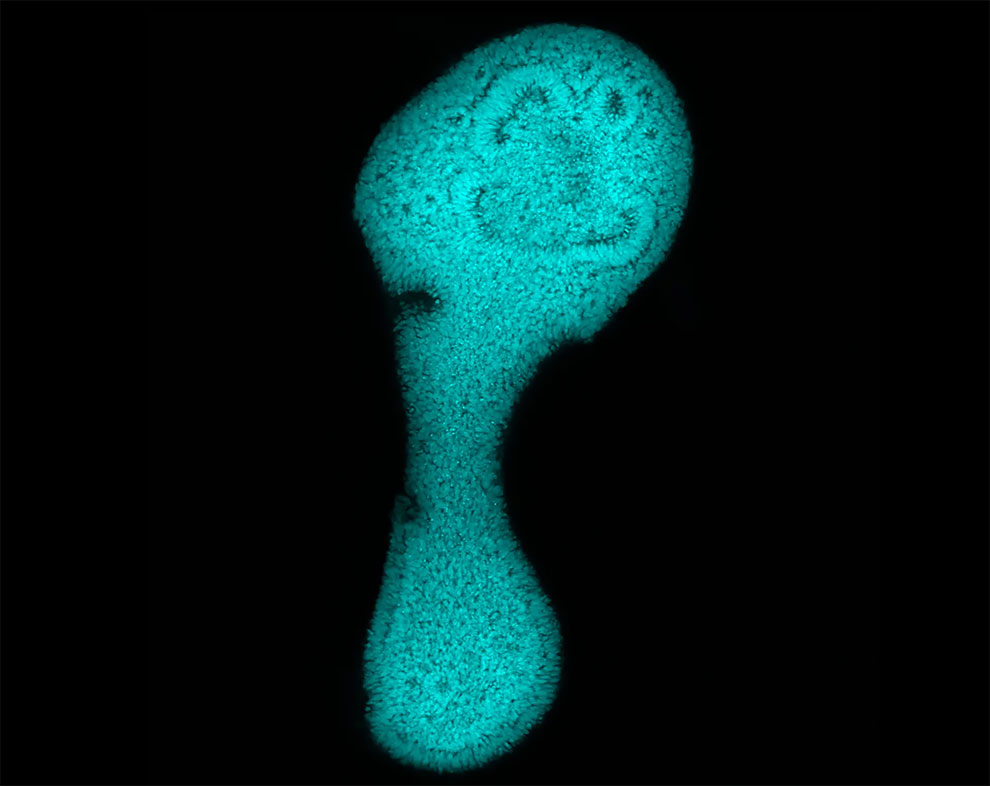
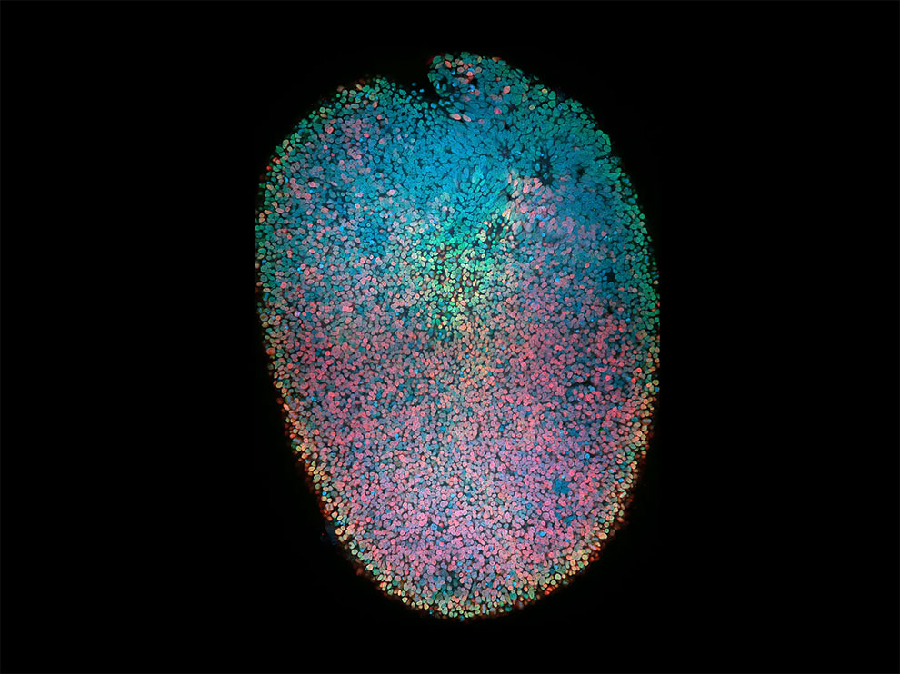
Hey that foot is smiling at us. Just kidding, it’s a self-organised axial organoid, cheekily exhibiting neural tube elongation. Photograph: Mohammed Shaker/AIBN
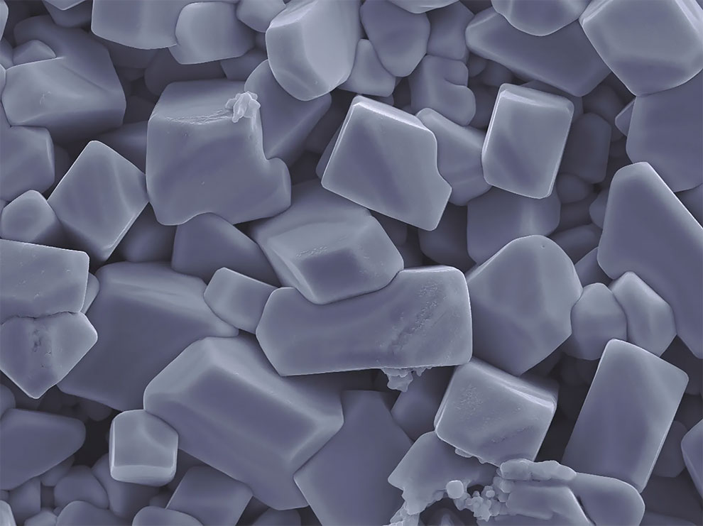
Pebbles? Milky quartz? The world’s smoothest Lego? Nope, these are structures of dissolved solar cells. Photograph: Yongxin Huang/AIBN
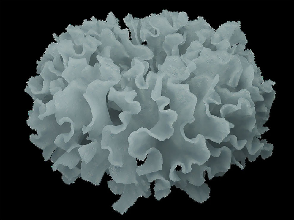
These cabbage-like nickel phosphate particles have been synthesised by a solvothermal process. Not suitable for Chiko Roll filler. Photograph: Valentino Kaneti/AIBN
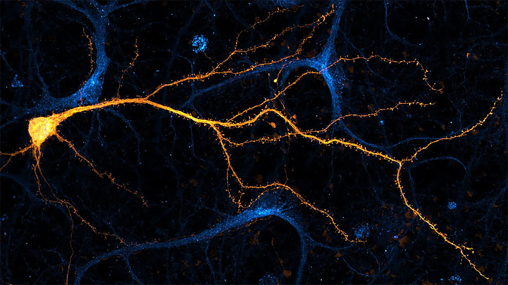
Here, a Diskovery spinning disk confocal microscope has been used to image cultured hippocampal neurons (the yellow bits) immunostained against endogenous lipid modifying enzyme DDHD2 (the blue bits). Photograph: Saber Abd Elkader/AIBN
Organoid science involves growing tiny synthetic copies of a person’s internal organs for drug and disease research. This human spinal cord organoid shows the segregation of cell populations that develop into neurons and motor neurons. Photograph: Sean Morrison/AIBN
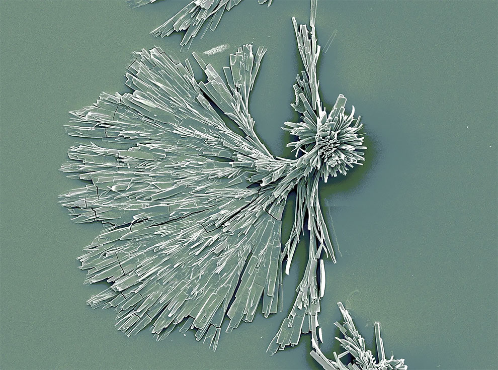
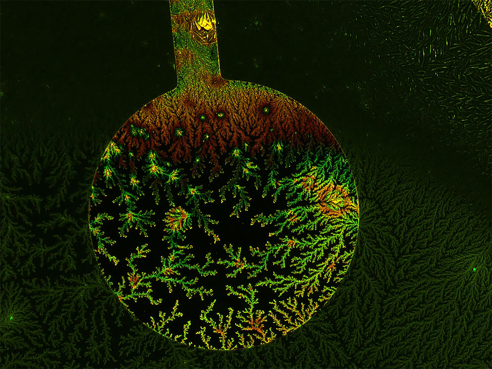
These carbon-based metal–organic framework nanorods have gradually decomposed and aggregated in a water solution, finally forming elegant radial crystals. Photograph: Ping Cheng/AIBN
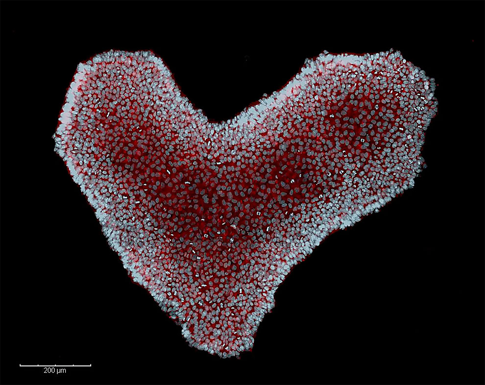
Stem cell science can sometimes make your heart skip a beat. This human embryonic stem cell colony consists of hundreds of cells clustered together in a familiar and lovely shape. Photograph: Desi Veleva/AIBN
Don’t let the purple hue fool you – this image captured by a scanning electron microscope is actually showing us mesoporous gold nanoparticles. Photograph: Javeria Bashir/AIBN
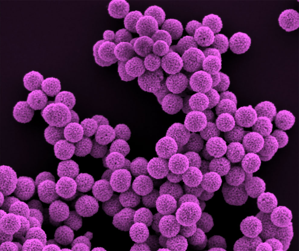
This image might appear festive, but we assure you it is not. This fluorescent microscope image captures the receptor binding domain of the Sars-CoV-2 spike protein. Photograph: Harshita Rupani/AIBN
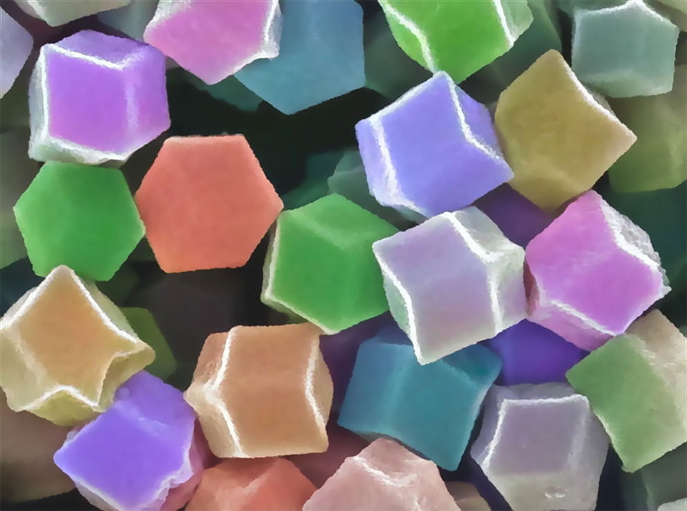
Here we have an image of some zeolitic imidazolate framework-8 rhombic dodecahedron particles. The technical term, we understand, is ‘science candies’. Photograph: Valentino Kaneti/AIBN
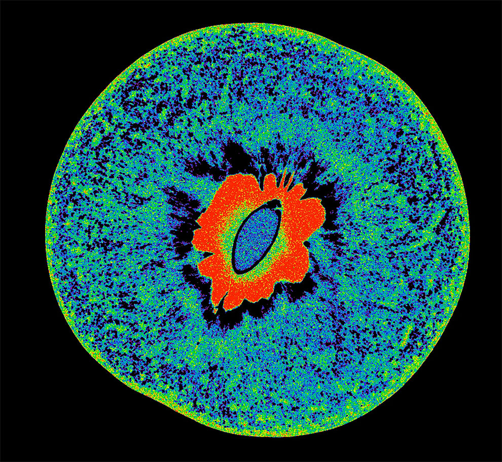
Sometimes you’ve just got to know what a piece of fruit looks like on the inside. To that end, here’s a juicy nectarine under microCT imaging. Photograph: Gary Cowin/AIBN